



Hearing that you may need brain surgery can bring up many questions. Two of the most common questions are how much of the skull needs to be opened, and how long the recovery will be after surgery. Answers to these questions depend heavily on your diagnosis. Some brain conditions require a traditional opening in the skull to give the surgeon direct access, while others can be treated through a smaller opening or with instruments guided through blood vessels. Experienced brain surgeons can also use advanced imaging to help navigate around delicate structures during an operation.
Craniotomy, endoscopic surgery, and catheter-based procedures are just some of the many ways brain surgery can be performed. Read on to learn the differences between these procedure types and where to find the best brain surgeon in Los Angeles to guide you through finding the right procedure for your needs.
What Is the Difference Between Open and Minimally Invasive Brain Surgery?
The main difference between these two surgical techniques is how the surgeon reaches the area that needs treatment:
Traditional Open Brain Surgery
Open brain surgery typically includes a procedure called a craniotomy. With this approach, the surgeon makes an incision in the scalp and temporarily removes a section of the skull to reach the brain. After completing the procedure, the surgeon replaces and secures the bone.
A craniotomy gives the surgeon room to see and work directly around the affected area. This approach is often recommended when a condition requires a large view, access to complex anatomy, or the removal of tissue that cannot be reached safely through a smaller opening. This can include conditions such as:
- Brain tumors that require surgical removal, including meningiomas, gliomas, and metastatic lesions
- Brain aneurysms that are better treated with microsurgical clipping
- Conditions that require direct access to relieve pressure or address an abnormality
- Complex cases where a smaller surgical corridor would limit safe access
Minimally Invasive and Keyhole Brain Surgery
Minimally invasive brain surgery uses a smaller opening or a narrow surgical route to reach a specific area. In some procedures, the surgeon uses an endoscope, a thin tube with a light and camera, along with small instruments.
Keyhole or endoscopic techniques are considered for select brain tumors and other conditions that are accessible through a narrow pathway. For example, some tumors near the skull base can be reached through the nasal passages, while certain deep-seated lesions may be approached through a small opening in the skull.
Potential benefits of minimally invasive or keyhole techniques include:
- A smaller incision or opening
- Less disruption to surrounding tissue in suitable cases
- A recovery plan that may involve less healing at the surgical access site
How Does Image-Guided Brain Surgery Work?
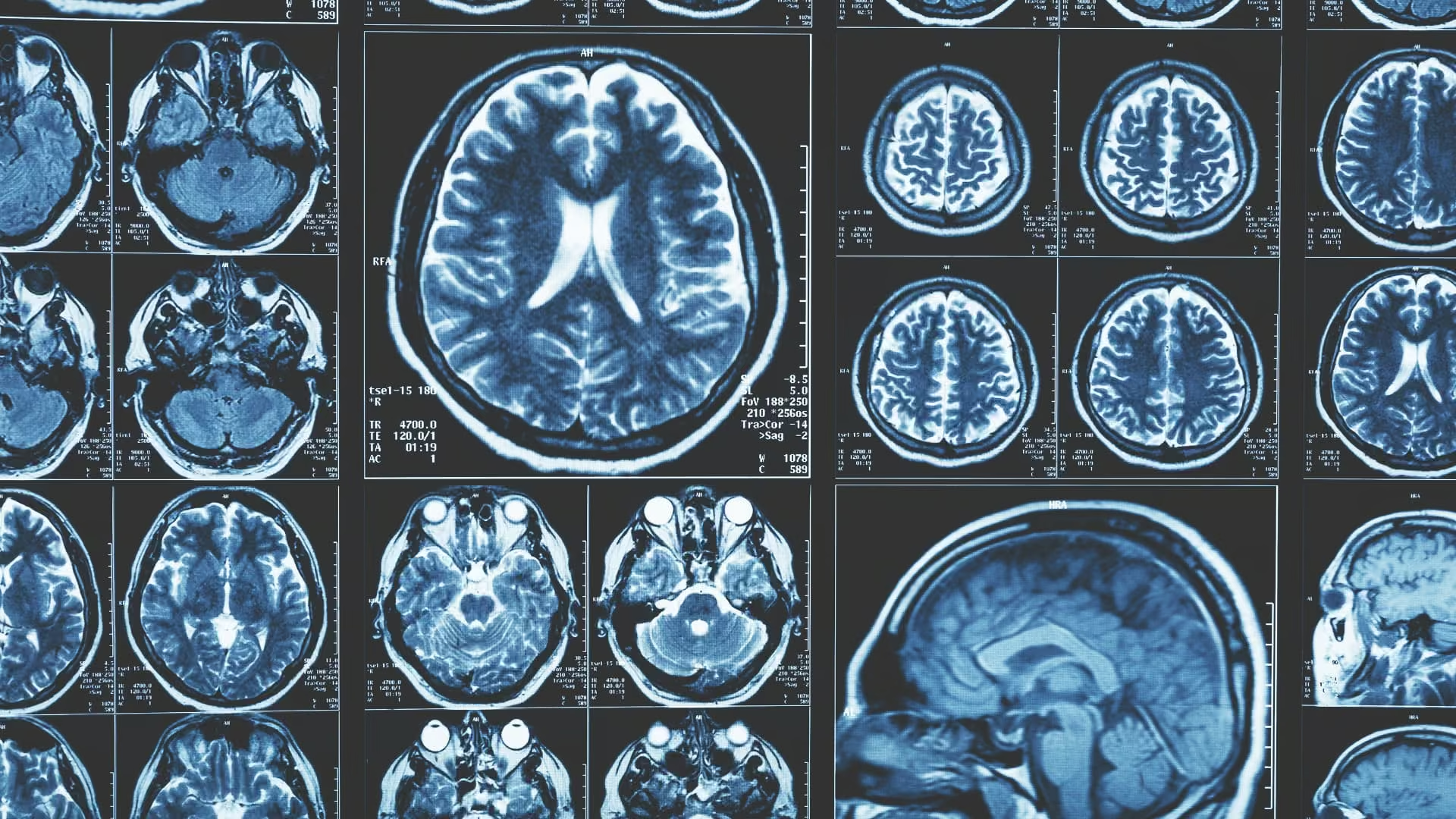
Image-guided surgery uses scans and computer-assisted navigation to help the surgeon locate the target during an operation. Before surgery, MRI or CT images can create a detailed map of the brain and the area that needs treatment. Then during the procedure, a specialized navigation system helps the surgeon understand where instruments are positioned in relation to the target and nearby structures. This is particularly helpful when a tumor or abnormality lies near areas responsible for movement, speech, vision, or other important functions.
For brain tumor surgery, image-guided navigation can help the surgeon:
- Locate the tumor and plan a path toward it
- Understand the tumor’s position in relation to nearby structures
- Guide instruments toward the intended area
- Limit unnecessary disruption to healthy tissue
Does Minimally Invasive Brain Surgery Mean a Faster Recovery?
A smaller incision may mean less healing at the access site, but it does not guarantee a shorter recovery. Recovery really depends on the procedure, the condition being treated, and your overall health.
Recovery after brain surgery is not something you’ll face alone. Your care team will monitor you for changes in neurological function and other complications. Your recovery plan may include follow-up imaging, medication, activity restrictions, or rehabilitation.
Which Brain Surgery Approach Is Right for You?
The safest brain surgery approach depends on the condition, its location, and its complexity. A tumor near the skull base may be accessible through the nose or a small opening, while another may require a craniotomy for safe access. Surgeons also consider nearby areas that control speech, movement, and vision, as well as the size and shape of the abnormality. The only way to know which brain surgery is right for you is with the guidance of a highly skilled expert.
Find the Best Brain Tumor Surgeon in Los Angeles for Your Needs
When brain surgery is on the horizon, getting the best treatment requires a thorough review of your diagnosis and imaging, along with a clear understanding of your treatment options.
Dr. Parham Yashar is a board-certified, fellowship-trained neurosurgeon who treats complex brain conditions, including brain tumors and aneurysms. His training in neurological surgery and endovascular neurosurgery allows him to consider microsurgical, minimally invasive, image-guided, and catheter-based approaches based on each patient’s condition.
Our goal is to always select an approach that addresses the condition while protecting healthy brain tissue and preserving neurological function. This is why Dr. Yashar always considers conservative treatment, like watchful observation or non-surgical options, when they can safely manage the condition.
Ready to learn which treatment approach may be right for you with help from the best brain surgeon in Los Angeles?

