


It is unsettling to hear the words “possible meningioma” after an MRI—especially if you were only looking for an explanation for headaches that feel different, a first seizure, new double vision, or numbness in your face or arm. One of the most helpful next steps is understanding how doctors describe types of meningioma. In most cases, “type” is less about a catchy name and more about where the tumor sits and which nearby nerves, blood vessels, or brain structures it may affect.
Below, we walk through common meningioma locations, the symptoms they may cause, how diagnosis is confirmed, and how treatment is typically considered. If you already have imaging, this will help you ask better questions during a specialist visit and understand the reasoning behind recommendations for meningioma treatment.
What Is a Meningioma?
A meningioma is a tumor that develops from the meninges, the thin protective layers covering the brain and spinal cord. Many meningiomas are benign (non-cancerous) and slow-growing, but some are atypical or malignant and can grow faster or recur more often.
Even when a meningioma is benign under the microscope, it can still cause significant symptoms if it presses on the brain, spinal cord, cranial nerves (which control vision, facial sensation, swallowing, and hearing), or major blood vessels. That is why two people with “the same diagnosis” can have very different experiences—and why the tumor’s location plays such a big role in treatment planning.
Common Symptoms of Meningioma
Meningioma symptoms depend on where the tumor is and which structures are being irritated or compressed. Some people have no symptoms at all, and the tumor is found incidentally during imaging for an unrelated problem. Others notice a gradual change that becomes harder to ignore.
Common symptoms include:
- Headaches that are new, persistent, or worsening
- Seizures (including a first-time seizure)
- Vision changes (blurred vision, double vision, loss of peripheral vision)
- Dizziness or balance problems
- Weakness, heaviness, or numbness in an arm or leg
- Facial numbness, tingling, or facial muscle weakness
- Memory, personality, or concentration changes (often with tumors near the frontal lobe)
- Difficulty walking or coordination problems
If you have sudden severe symptoms—such as a first-time seizure, sudden weakness on one side, confusion, or rapid vision loss—seek urgent medical care.
Why “Type” Often Means Location (and Why It Matters)
Meningiomas can be categorized by tumor grade (based on cell features under a microscope), but in day-to-day care, many discussions focus on anatomical location. Location helps your specialist estimate:
- Which symptoms are most likely to be related to the tumor
- How close the tumor is to critical nerves and blood vessels
- Whether complete removal is feasible and safe
- When monitoring or radiation may be preferred over surgery
The sections below describe common meningioma locations and the practical issues each can raise.
Types of Brain Meningiomas by Location
Convexity Meningioma
Convexity meningiomas grow on the outer surface of the brain, just beneath the skull. Because they are not deep within the skull base, they are often more straightforward to access than tumors surrounded by cranial nerves and major arteries.
Symptoms depend on the nearby brain region and can include headaches, seizures, or weakness and numbness on one side of the body.
Falcine (or Parasagittal) Meningioma
Falcine and parasagittal meningiomas arise near the falx, a fold of tissue that separates the two hemispheres. Important veins and drainage pathways may be nearby, so surgical planning often focuses on minimizing risk to those structures.
Depending on exact position, symptoms may include seizures, changes in sensation, or weakness—sometimes more noticeable in the leg than the arm.
Intraventricular Meningioma
Intraventricular meningiomas occur within the ventricles, the fluid-filled spaces that contain cerebrospinal fluid (CSF). If they partially block CSF flow, they can contribute to pressure buildup inside the skull.
Symptoms may include headaches, nausea, dizziness, or signs related to hydrocephalus when obstruction is significant.
Skull Base Meningiomas (Near Vision, Eye Movement, and Facial Nerves)
Skull base meningiomas develop along the bottom portion of the skull, where cranial nerves and major blood vessels travel through narrow spaces. Because the anatomy is dense in this region, symptoms often involve vision, eye movement, facial sensation, swallowing, or hearing. Treatment is typically individualized and may include observation, surgery, radiation, or a combination.
Sphenoid Wing Meningioma
Sphenoid wing meningiomas grow near the bone behind the eye. Their proximity to the optic nerve and the nerves controlling eye movement means symptoms may develop gradually, such as trouble reading, bumping into objects on one side, double vision, or new eye strain.
Depending on the tumor’s extent, care may involve careful observation, surgery, radiation, or combined strategies to balance tumor control with nerve protection.
Cavernous Sinus Meningioma
The cavernous sinus contains cranial nerves responsible for eye movement and facial sensation, along with critical blood vessels. Meningiomas in this area may cause double vision, drooping eyelid, facial numbness or pain, or difficulty moving the eye normally.
Because complete surgical removal can risk nerve injury in some cases, specialists often weigh multiple options, including observation or focused radiation when appropriate.
Suprasellar Meningioma
Suprasellar meningiomas are located near the pituitary region and the optic apparatus. Visual symptoms are common, including loss of peripheral vision or reduced sharpness in one or both eyes.
Evaluation typically includes a careful eye exam and MRI review to understand how the tumor relates to the optic nerves and nearby blood vessels.
Clival Meningioma
Clival meningiomas develop near the clivus, a bone at the skull base close to the brainstem and several cranial nerves. Symptoms vary with growth pattern and can include headaches, facial symptoms, swallowing changes, or coordination issues.
Because approaches to this area can be complex, detailed imaging review is central to choosing a treatment plan.
Foramen Magnum Meningioma
Foramen magnum meningiomas arise at the junction where the brain transitions into the spinal cord. This compact region carries vital pathways, so even relatively small tumors can cause symptoms such as neck pain, weakness, numbness, balance issues, or changes in walking.
When symptoms are progressive, timely specialty evaluation matters to prevent worsening neurological function.
Tentorial Meningioma
Tentorial meningiomas grow along the tentorium, a membrane that separates the cerebrum from the cerebellum. Symptoms depend on nearby structures and may include headaches, seizures, or balance and coordination problems.
Because this region sits near important venous structures, the safest treatment path depends on anatomy seen on imaging.
Spinal Meningiomas
Meningiomas can also develop along the spinal meninges. Spinal meningiomas may initially feel like a “spine problem” rather than a tumor, especially when symptoms come on gradually.
Symptoms can include:
- Progressive weakness or heaviness in the legs
- Numbness or tingling below a certain level
- Changes in walking, balance, or coordination
- Pain that wraps around the chest or abdomen (depending on the spinal level)
Because spinal tumors can compress the spinal cord, persistent or progressive symptoms deserve prompt evaluation. For related symptoms and differential diagnoses, you can also review our spine conditions hub.
How Meningiomas Are Diagnosed
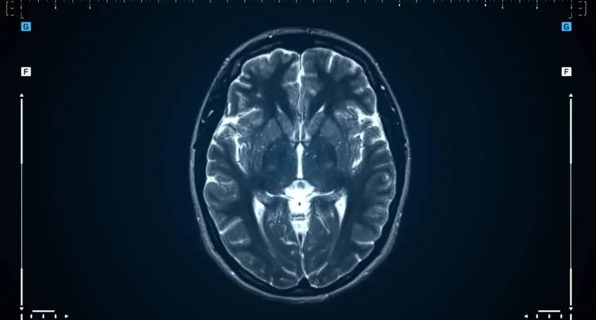
Diagnosis usually begins with imaging—most often an MRI, which shows soft tissues in detail and helps define the tumor’s relationship to the brain, spinal cord, cranial nerves, and blood vessels. A CT scan may be added to evaluate how the tumor interacts with bone or to clarify calcification.
Imaging helps estimate size, location, and features that suggest growth behavior, but tumor grade is often confirmed only after tissue is obtained—commonly at the time of surgery.
Treatment Options for Meningioma
Treatment is based on symptoms, tumor size and location, evidence of growth on serial imaging, overall health, and your goals. Many plans involve one of the following approaches:
Observation (Watchful Waiting)
For small, slow-growing tumors that are not causing symptoms, observation with scheduled imaging can be appropriate. This can be a thoughtful choice when a tumor is stable and the risks of intervention outweigh the benefits at that time.
Surgery
Surgery aims to remove as much tumor as safely possible and relieve pressure on surrounding structures. Whether complete removal is realistic depends heavily on location. When a tumor involves delicate cranial nerves or major vessels, the safest plan may be partial removal with additional therapy to control the remainder.
If surgery is part of your plan, you can explore the practice’s brain surgery resources, including meningioma surgery. In select cases, a minimally invasive brain tumor surgery approach may be discussed, depending on tumor location and anatomy.
Radiation Therapy
Radiation may be used when surgery is not the safest option, when residual tumor remains after surgery, or when imaging suggests a higher likelihood of regrowth. The goal is tumor control—slowing or stopping growth—while protecting surrounding tissue.
Medications for Symptom Control
Medications do not remove a meningioma, but they can help manage symptoms. Anti-seizure medication may be used if seizures occur, and other medicines may be recommended to address swelling or headache patterns based on your clinical situation.
When to See a Neurosurgeon (and When to Get a Second Opinion)
Consider a neurosurgical evaluation if you have been diagnosed with a meningioma, your imaging report suggests one, or you have neurologic symptoms that are persistent or worsening.
A second opinion can be helpful when:
- The tumor is at the skull base or near the optic nerves
- You have symptoms that do not seem to match the imaging explanation
- You were told surgery is the only option without discussing monitoring or radiation
- You want clarity on the risks of observation versus intervention
Learning more about broader brain tumor treatment options can also help you prepare for these discussions.
Finding the Best Meningioma Surgeon in Los Angeles
Meningiomas can sit next to structures that control vision, facial movement, balance, and strength. When treatment is needed, the details matter—exact tumor location, blood vessel relationships, and how to relieve pressure while protecting function.
If you are looking for the best meningioma surgeon in Los Angeles, Parham Yashar, MD and the team at Yashar Neurosurgery provide careful, patient-centered evaluation and clear explanations of your options, including observation, surgery, and adjunct therapies when appropriate. To discuss symptoms, review imaging, or request a second opinion, call (424) 209-2669 or schedule a consultation at our office at 8436 W. 3rd Street, Suite 800, Los Angeles, CA 90048.
