


Seeing “aneurysm” on an imaging report can make everything else fade into the background. Patients commonly ask the same urgent questions: What kind is it? Does the type change the risk? And does it mean I need treatment right away? Understanding the types of brain aneurysms can help you make sense of the language in your report and why one person is monitored while another is treated.
Aneurysm care is not one-size-fits-all. Specialists look at the aneurysm’s shape, neck, size, and location, along with your symptoms and health history, to estimate risk and recommend the most appropriate next step. For an overview of how aneurysms are evaluated and managed, you can also visit our page on brain aneurysm treatment.
What a Brain Aneurysm Is (and Why Type Matters)
A brain aneurysm is a weak or thin area in the wall of an artery in the brain that bulges outward as blood flows through it. Many aneurysms are found incidentally during imaging for unrelated symptoms, and many never rupture. The concern is that an aneurysm can leak or rupture and cause bleeding around the brain (subarachnoid hemorrhage), which is a medical emergency.
“Type” is shorthand for how an aneurysm forms and what it looks like. That matters because different shapes behave differently, and some are better suited to endovascular treatment (through the blood vessels) while others may be better treated with microsurgery (open surgery). Type is only one piece of the puzzle, but it’s an important one.
Types of Brain Aneurysms
Doctors describe aneurysms by their shape and cause. Below are the major categories you may see in a report, plus what they can mean clinically.
Saccular (Berry) Aneurysms
Saccular aneurysms are the most common type and are often called “berry aneurysms” because they look like a small rounded pouch arising from a blood vessel. In the source draft, they are described as accounting for about 80% to 90% of brain aneurysms. They often form at branching points of arteries, where blood flow forces can be higher.
- Shape: Rounded dome with a more defined neck
- Typical location: Branch points in the arteries at the base of the brain
- How they present: Often silent until they enlarge, irritate nearby nerves, leak, or rupture
If an unruptured saccular aneurysm presses on nearby nerves, symptoms can include headaches, eye pain, drooping eyelid, or vision changes. Many other conditions can cause these symptoms, but a new or worsening pattern should be evaluated.
Fusiform Aneurysms
Fusiform aneurysms involve a widening of the artery itself rather than a single pouch. The vessel bulges around its circumference, and there is usually no distinct neck. These aneurysms are often associated with underlying vessel disease such as atherosclerosis.
- Shape: Spindle-like widening of a segment of artery
- Neck: Typically absent (unlike saccular aneurysms)
- Common location: Can occur in larger arteries, including those in the back of the brain (vertebrobasilar system)
Fusiform aneurysms may cause symptoms by affecting blood flow or compressing nearby structures rather than rupturing. Depending on the artery involved, symptoms can include dizziness, vision changes, imbalance, or coordination difficulty.
Dissecting Aneurysms
A dissecting aneurysm occurs when there is a tear in the inner lining of an artery. Blood can track between layers of the vessel wall, which may narrow the artery, disrupt normal blood flow, and weaken the vessel. Dissections can be associated with trauma to the head or neck, connective tissue disorders, or spontaneous vessel wall degeneration.
- Who it can affect: Can occur in younger patients compared with degenerative aneurysms
- Why it matters: Can cause stroke-like symptoms if blood flow is reduced or clots form
- Risk: Varies by location and severity of the dissection
Possible symptoms include sudden severe headache, neck pain, facial droop, weakness, numbness, or difficulty speaking. Sudden neurologic symptoms should be treated as an emergency.
Mycotic (Infectious) Aneurysms
Mycotic aneurysms are caused by infection that damages and weakens an artery wall. Despite the name, they are often related to bacterial infection, including infective endocarditis (infection involving the heart valves) that can spread through the bloodstream.
- Cause: Infection-related weakening of the vessel wall
- Appearance: May be irregularly shaped and sometimes multiple
- Clinical issue: Vessel walls can be fragile, raising concern for rupture
Symptoms can include fever or chills along with neurologic symptoms, depending on location. Management often requires coordination of infection treatment and aneurysm stabilization.
Traumatic Aneurysms
Traumatic aneurysms develop after a head injury damages a blood vessel. They are uncommon, but the risk of bleeding can be higher because the vessel wall is directly injured. Some develop soon after trauma, while others appear later.
- Cause: Direct vessel injury from head trauma
- Shape: Often irregular with fragile walls
- Presentation: Can show up with headache, seizures, confusion, or bleeding depending on severity
Warning Signs and When to Seek Emergency Care
Many aneurysms do not cause symptoms. Symptoms may occur when an aneurysm grows, presses on nearby nerves, leaks, or ruptures.
Call 911 or go to the ER immediately for symptoms concerning for a ruptured aneurysm, including a sudden severe headache (often described as the worst headache of one’s life), fainting, seizures, confusion, sudden weakness or numbness, or severe neck stiffness.
Symptoms that can sometimes be linked to an unruptured aneurysm (depending on its size and location) include new or unusual headaches, vision changes, drooping eyelid, facial numbness, or eye pain. These symptoms have many possible causes, but they should be evaluated promptly when they are sudden, worsening, or paired with neurologic changes.
Risk Factors and Potential Complications
Although aneurysm types differ, several factors are associated with a higher likelihood of developing an aneurysm or having complications.
Risk Factors
- Family history of brain aneurysms
- High blood pressure, which can stress artery walls over time
- Smoking, which damages blood vessels and increases rupture risk
- Age and sex, with higher prevalence in adults and particularly in women
- Stimulant use (such as cocaine), which can cause sudden blood pressure spikes
Complications
The most serious complication is rupture, which can lead to hemorrhagic stroke, brain injury, or death. Unruptured aneurysms can still cause problems if they compress nearby nerves or brain tissue or alter blood flow in ways that raise the risk of clotting or stroke.
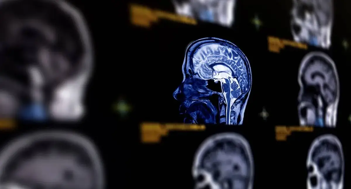
How Brain Aneurysms Are Diagnosed
Diagnosis relies on imaging, and the right test depends on the situation. If a ruptured aneurysm is suspected, the focus is on quickly identifying bleeding and finding the source. If an aneurysm is found incidentally, the goal is to define its anatomy well enough to estimate risk and plan next steps.
- CT scan: Often used to rapidly detect bleeding in and around the brain
- MRI: Provides detailed imaging of brain structures and surrounding tissue
- Cerebral angiography: Evaluates blood flow and vessel anatomy in high detail for planning treatment
When an aneurysm is identified, specialist review focuses on details that affect safety and options, such as neck width, branch vessels near the aneurysm, and whether the artery wall looks diseased or dissected.
Treatment Options: Monitoring, Endovascular Therapy, and Microsurgery
Treatment depends on whether the aneurysm is ruptured, the aneurysm’s type and anatomy, and your personal risk profile. Some small unruptured aneurysms may be monitored with periodic imaging, while others are treated proactively to reduce rupture risk.
- Monitoring: Selected small, unruptured aneurysms may be followed with imaging and risk-factor management (such as blood pressure control and smoking cessation)
- Neuroendovascular coiling: A minimally invasive approach performed through the blood vessels to fill the aneurysm and reduce blood flow into it; learn more about neuroendovascular coiling
- Flow diversion: Uses a specialized stent to redirect blood away from the aneurysm to promote healing of the vessel wall in appropriate cases; see flow diversion
- Microsurgical clipping: An open surgery that places a clip at the aneurysm’s base to stop blood from entering it; read about microsurgical clipping
Some aneurysms need additional support to treat the aneurysm while preserving normal blood flow through nearby branches. Techniques can include stent-assisted coiling or balloon-assisted coiling.
Finding the Best Neuroendovascular Surgeon in Los Angeles
If you’ve been told you have an aneurysm—or your report lists a term like “saccular,” “fusiform,” or “dissecting”—a specialist evaluation can turn unfamiliar wording into a clear plan. A helpful consultation typically includes a careful review of your imaging, an explanation of why your aneurysm’s type and anatomy matter, and a straightforward discussion of the options: monitoring, endovascular treatment, or microsurgery.
At Yashar Neurosurgery, Parham Yashar, MD provides individualized aneurysm evaluation and treatment planning, including advanced endovascular approaches and microsurgical techniques when appropriate. You can explore related services in our brain conditions and brain surgery pages.
If you need an expert opinion on a newly diagnosed aneurysm, changing symptoms, or treatment options, contact Yashar Neurosurgery in Los Angeles at (424) 209-2669 or request a consultation at 8436 W. 3rd Street, Suite 800, Los Angeles, CA 90048.
